Home
Conferences
Cases
About
Contact
Sign In
Breast
Chest
Brain
Head and Neck
Gastrointestinal
Genitourinary
Heart
Skeletal
Spine
Vasculature
Neuroradiology Medical Terminology
Corona Virus
YouTube Lectures
e Books
Radiology Links and Databases
Online Webinars
Tips and Tricks in Imaging
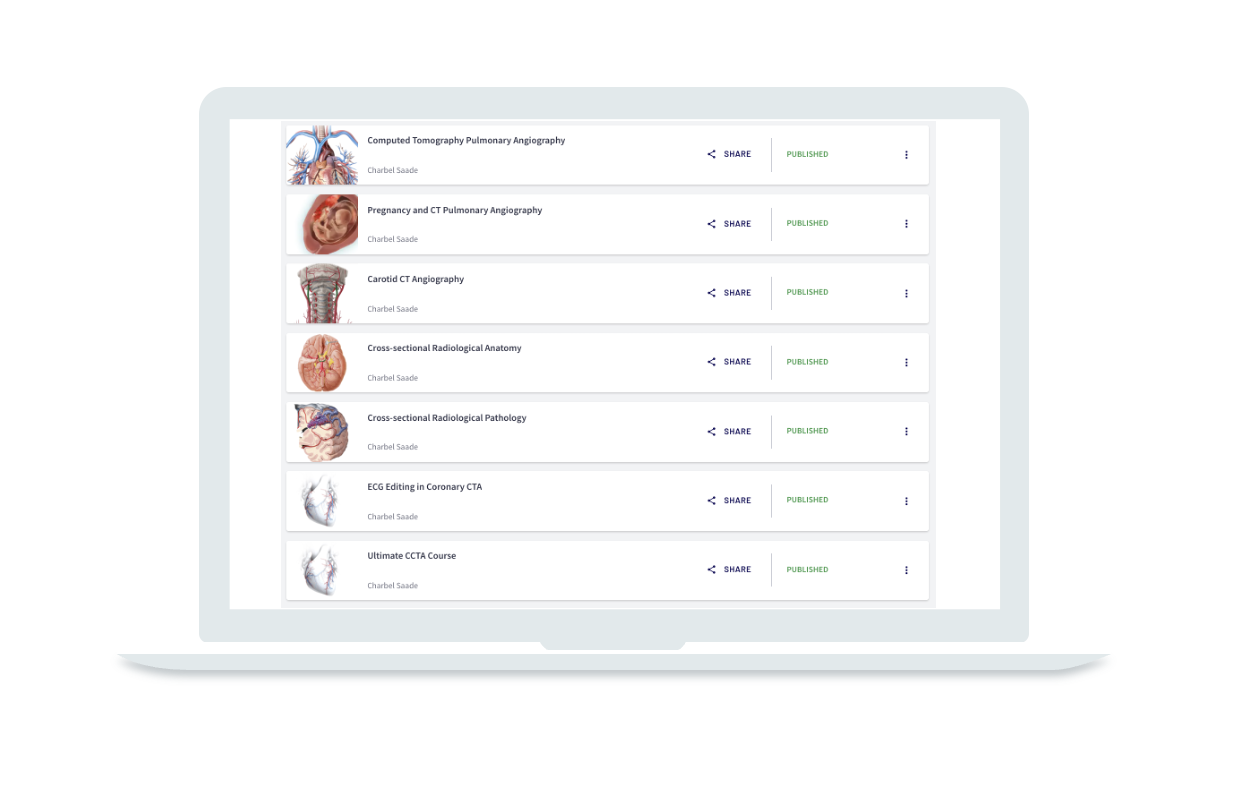
Online Courses